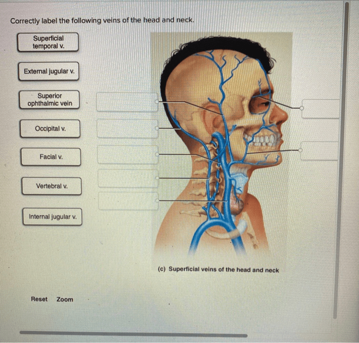
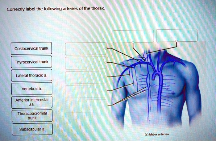
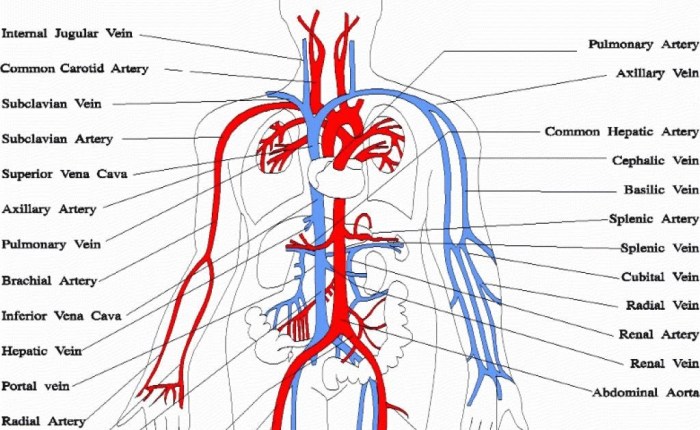
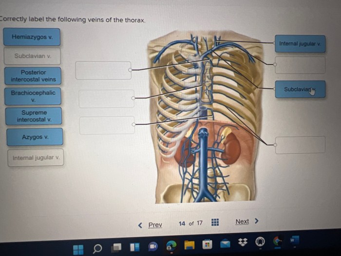
Correctly label the following arteries of the thorax – Correctly labeling the arteries of the thorax is paramount for a thorough understanding of human anatomy. This article provides a comprehensive guide to accurately identifying and understanding the significance of these vital structures.
The thoracic arteries, originating from the aorta, play a crucial role in supplying oxygenated blood to the heart, lungs, and other thoracic organs. Mislabeling or misinterpreting these arteries can lead to severe consequences, emphasizing the importance of accurate labeling.
Introduction

The thoracic arteries are a complex network of vessels that supply oxygenated blood to the heart, lungs, and other structures within the chest cavity. Understanding the anatomy of these arteries is crucial for various medical procedures and diagnostic tests. This article aims to provide a comprehensive guide to correctly labeling the arteries of the thorax.
Anatomy of the Thoracic Arteries
Aorta
The aorta is the largest artery in the body, originating from the left ventricle of the heart. It descends through the chest cavity, giving off branches that supply blood to the head, neck, arms, and internal organs.
Brachiocephalic Trunk
The brachiocephalic trunk is the first major branch of the aorta. It divides into the right common carotid artery and the right subclavian artery, which supply blood to the right side of the head, neck, and arm.
Common Carotid Artery
The common carotid arteries, left and right, ascend through the neck, giving off branches that supply blood to the brain and face.
Subclavian Artery
The subclavian arteries, left and right, supply blood to the arms and shoulders. They give off branches that nourish the muscles, bones, and skin of the upper extremities.
Internal Thoracic Artery, Correctly label the following arteries of the thorax
The internal thoracic arteries, left and right, arise from the subclavian arteries. They run along the inner surface of the chest wall, supplying blood to the breast and abdominal wall.
Pericardiophrenic Artery
The pericardiophrenic arteries arise from the internal thoracic arteries. They supply blood to the pericardium, the sac surrounding the heart, and the diaphragm.
Bronchial Arteries
The bronchial arteries, usually two in number, arise from the aorta. They supply blood to the lungs, providing oxygen and nutrients for respiration.
Esophageal Arteries
The esophageal arteries arise from the aorta or the left gastric artery. They supply blood to the esophagus, the muscular tube that connects the throat to the stomach.
Clinical Significance of the Thoracic Arteries

Accurately labeling the thoracic arteries is crucial for various medical procedures and diagnostic tests. Mislabeling or misinterpreting these arteries can lead to incorrect diagnoses, ineffective treatments, and potential complications. For instance, during cardiac catheterization, precise identification of the coronary arteries is essential for successful angioplasty or stenting procedures.
Methods for Labeling the Thoracic Arteries

Dissection
Dissection involves physically exposing the arteries through surgical techniques. This method provides a direct view of the arteries, allowing for detailed examination and labeling. However, it is invasive and carries risks associated with surgery.
Angiography
Angiography involves injecting a contrast agent into the arteries and using X-rays to visualize their structure and course. It is a minimally invasive technique that provides detailed images of the arteries, but it exposes patients to radiation.
Magnetic Resonance Imaging (MRI)
MRI uses strong magnetic fields and radio waves to create cross-sectional images of the body. It can visualize the thoracic arteries non-invasively, but it may not provide as much detail as angiography.
Computed Tomography (CT)
CT scans use X-rays and computer processing to create detailed cross-sectional images of the body. It can visualize the thoracic arteries, but it exposes patients to radiation and may not provide as much detail as angiography.
Tips for Correctly Labeling the Thoracic Arteries: Correctly Label The Following Arteries Of The Thorax

- Use a comprehensive anatomical atlas or reference book for accurate identification.
- Consult with experienced anatomists or radiologists for guidance.
- Utilize multiple imaging modalities, such as angiography and MRI, to cross-reference and confirm labeling.
- Pay attention to the branching patterns, relationships with surrounding structures, and the course of the arteries.
- Avoid relying solely on visual cues or preconceived notions, as variations in anatomy can occur.
- Ensure proper patient positioning and image orientation during imaging procedures to minimize distortion or misalignment.
Expert Answers
What is the purpose of labeling the arteries of the thorax?
Accurate labeling aids in understanding their anatomy, clinical significance, and guiding medical procedures.
What are the potential consequences of mislabeling thoracic arteries?
Mislabeling can lead to incorrect diagnosis, inappropriate treatment, and surgical complications.
What methods are used to label thoracic arteries?
Common methods include dissection, angiography, MRI, and CT scans.
